Published on January 2, 2018 - LinkedIn
As recently posted by the National Multiple Sclerosis Society, the McDonald Criteria for the Diagnosis of Multiple Sclerosis have just been revised by a 30-member international panel of MS experts. The team published their findings in The Lancet Neurology online on December 21, 2017. The entire contents of the paper can be found here.
The following changes, among others, were made: brain MRI should be obtained during the MS diagnostic process, unless not possible. Spinal MRI should be obtained when additional data are needed to confirm the diagnosis.
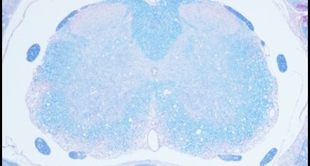
One of our students currently quantifies the proportion of MS imaging studies analyzing brain lesions versus spinal cord lesions. Not surprisingly, he found that most imaging and pathological MS studies are performed in the forebrain. By contrast, most EAE studies focus on spinal cord lesions.
Being a neuroanatomist, I am fully convinced that the spinal cord is not just a “simple” part of the brain, but fundamentally different from supratentorial parts of the central nervous system (CNS). Translation from bench to bedside might be hampered because of the discrepancy between regions of interest analyzed in pre-clinical and clinical MS studies. Of note, our lab recently developed a modified EAE protocol which leads to severe supratentorial inflammatory lesion development. This model will be used in the future to study underlying mechanisms of lesion development, lesion progression, and remyelination.
