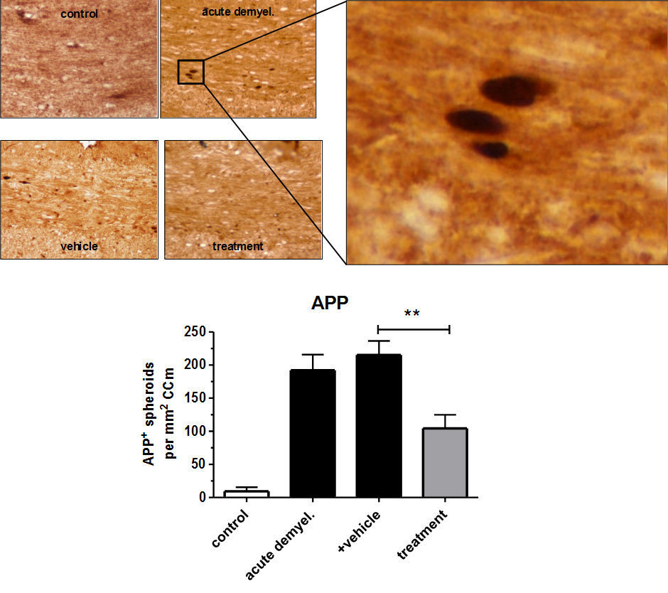
Acute axonal damage in the white matter tract corpus callosum is visualized by means of immunohistochemistry against amyloid precursor proteins (APP), a commonly used marker for disturbed axonal integrity. In this study, numbers of APP+ spheroids are dramatically lower in verum-treated compared to vehicle-treated mice. Specifically, 215 spheroids per mm2 were detectable in vehicle-treated animals, whereas 104 spheroids per mm2 were detectable in verum-treated animals (p=0.01). Thus, ongoing axonal damage was effectively suppressed by this pharmacological intervention.
Neuroprotection models
Protecting demyelinated axons is critical to maintain neural integrity as transection/loss of axons is the major cause of neurological decline in MS patients. The extent of axonal damage can be studied in various animal models.
Axonal transection or loss is a notable pathology in MS as a consequence of demyelination of axons in the brain. Protecting demyelinated axons is critical to maintain neural integrity as transection/loss of axons is the major cause of neurological decline in MS patients. ProMyelo’s mouse models can evaluate neuroprotection potential
of pharmaceutical compounds and biologics by analyzing the swollen and transected axons that are reminiscent of neuronal damage. In addition, ProMyelo’s electron microscopy capabilities allow comprehensive analysis of the neuroprotective effect by studying neuronal ultrastructural changes.
Experimental End Points
- Quantification of axonal damage (identified by axonal ovoids)
- Evaluation of survival/health of axons by immunohistochemistry
- Electron micoscopy evaluation of ultrastructural changes in axons
- Evaluation of mitochondrial integrity and changes