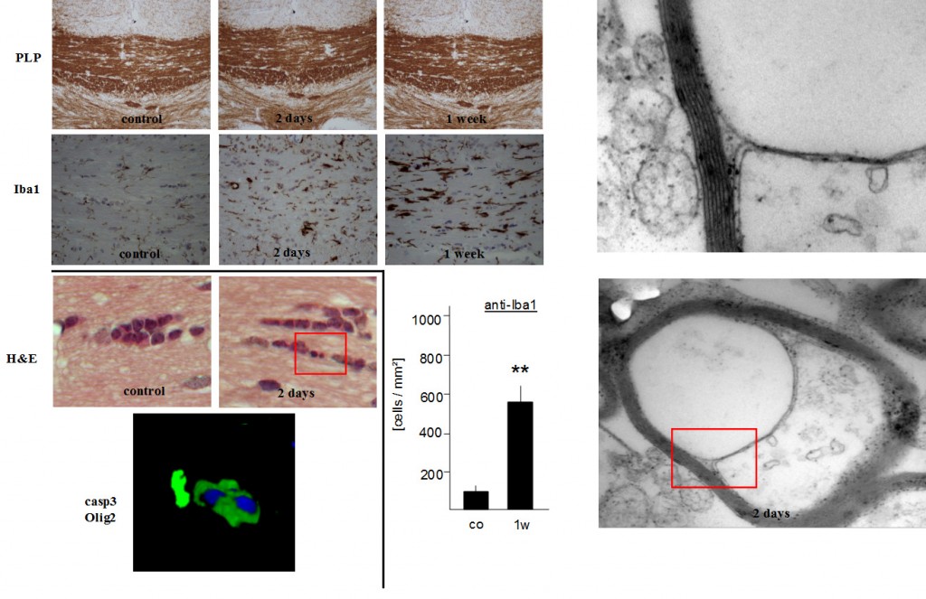
Characterization of early oligodendrocyte stress in cuprizone-treated mice. Numerous apoptotic oligodendrocytes with condensed and/or fragmented nuclei are seen as early as two days after cuprizone feeding (left red box). Oligodendrocyte apoptosis is paralleled by microglia activation. Ultrastructural analysis reveals signs of a dying back oligodendrogliopathy with a splitting of the inner myelin lamella (right red box) in the corpus callosum.
Demyelination models
Characteristic for Multiple Sclerosis lesions is the loss of myelinating oligodendrocytes and demyelination. ProMyelo’s modified cuprizone model shows consistent levels of axonal demyelination providing an excellent platform for qualitative and quantitative readouts of demyelination.
Cuprizone intoxication is one of several animal models used to study demyelination. Early treatment protocols exposed mice to cuprizone for 6 weeks to induce demyelination; however, more recent reports have varied exposure times from 4 to 5 weeks. We found that an abbreviated insult of only 3 weeks of exposure to cuprizone induces oligodedrocyte apoptosis with significant and reproducible demyelination 2 weeks later (5-week time point).
Once mature oligodendrocytes are perturbed after a 3-week treatment, the progression to demyelination occurs without requiring further exposure. This short, “hit and run” model triggers a cascade of events leading to demyelination 2-3 weeks later and is an excellent model to study the development of new therapeutic strategies for treatment of multiple sclerosis.
Experimental End Points
- Quantification of myelination in diverse brain regions
- Quantification of apoptotic oligodendrocytes
- Quantification of oligodendrocyte stress
- Additional histological quantification (OPCs, astrocytes, microglia, etc)
- Quantification of myelin protein expression levels
- Electron microscopy evaluation of distinct myelin and axonal parameters